Knee Tendon Diagram / Schematic Of The Knee Model With Contact Conditions And 21 Ligament Download Scientific Diagram : Jumper's knee is inflammation of your patellar tendon, the tendon that connects your kneecap (patella) to your shin bone (tibia).
Knee Tendon Diagram / Schematic Of The Knee Model With Contact Conditions And 21 Ligament Download Scientific Diagram : Jumper's knee is inflammation of your patellar tendon, the tendon that connects your kneecap (patella) to your shin bone (tibia).. Some of the most common symptoms of a torn knee ligament are pain, swelling and, in some cases, an audible snap. Tendons are the connection between bones and muscles. Bones, cartilage, ligaments, and tendons. When there is damage to one of the structures that surround the knee joint, this can lead to discomfort and disability. The kneecap slides along a groove in the femur as the knee bends.
The kneecap slides along a groove in the femur as the knee bends. It needs to be repaired and rehabilitated, says. Bones, cartilage, ligaments, and tendons. Then next one, further down, looks at pain behind the knee. Related posts of knee tendon anatomy diagram and name chart cross section of foot nerves.
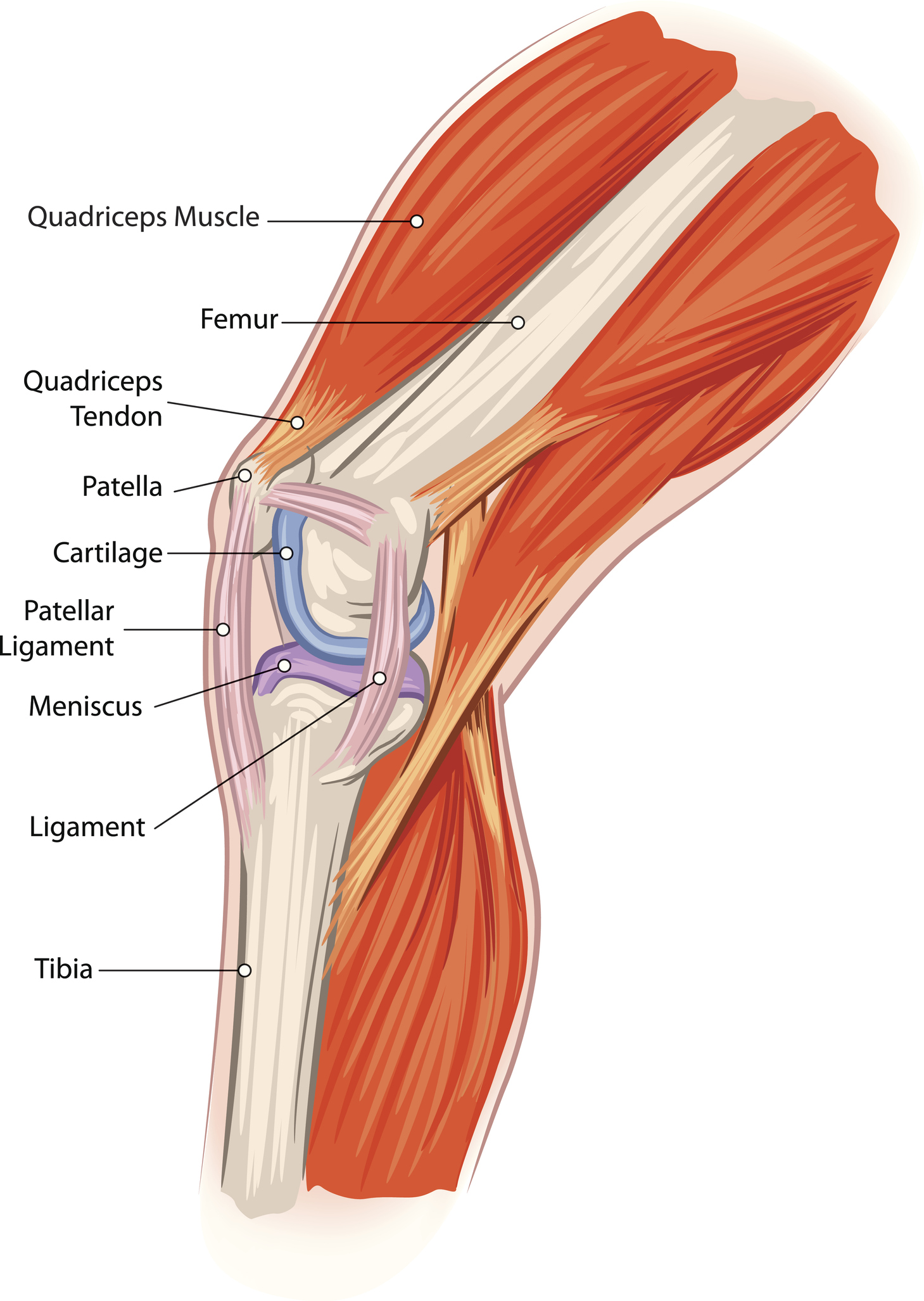
Patella Tendon Rupture Core Em from coreem.net Torn, weak, or stretched ligaments or tendons: The ligament, located in the center of the knee, that controls rotation. Vectorized and colorized in inkscape, based on image:knee diagram.png. Mcl & lcl found either side of the knee. You may be experiencing knee pain and want to know the possible causes. The knee joint is a complex structure that involves bones, tendons, ligaments, muscles, and other structures for normal function. Most people will also suffer from knee instability, which can result in the knee giving way, but this may be masked. Pain above the knee cap (yellow).
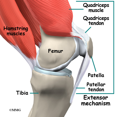
Your thighbone (femur), shinbone (tibia), and kneecap (patella).
The four main ligaments in the knee connect the femur (thighbone) to the tibia (shin bone), and include the following: Diagram of the ankle bones. Jumper's knee is inflammation of your patellar tendon, the tendon that connects your kneecap (patella) to your shin bone (tibia). Mcl & lcl found either side of the knee. Diagram of inside the body. There are numerous tendons around the knee that also help to stabilize the knee. Knee pain could be the result of a problem with any one of these components, or a combination of several. Three bones meet to form your knee joint: This svg file contains embedded text that can be translated into your language, using any capable svg editor, text editor or the svg translate tool. Cross section of foot nerves 13 photos of the cross section of foot nerves cross section of nerve fiber, foot anatomy nerves, foot nerve pain, human foot nerves, nerve cross section histology, peripheral nerve cross section, spinal nerve cross section, foot, cross section of nerve fiber, foot anatomy. One of the most important tendons is the. The knee is the largest joint in the body, and one of the most easily injured. The largest tendon in the knee is the patellar tendon which covers the kneecap runs up the thigh and attaches to the quadriceps.
Acl & pcl found in the middle of the joint. The severity of these symptoms depends on which ligament has been torn. This first knee pain diagnosis chart focuses on pain at the front of the knee. Its complexity and its efficiency is the best example of god's creation. The knee is the largest joint in the body who function is to bend (flex) and straighten (extend) in order to allow movement of the body e.g.
Knee Anatomy Eorthopod Com from eorthopod.com The knee is designed to fulfill a number of functions: Ligaments are elastic bands of tissue that connect bones to each other and provide stability and strength to the joint. It needs to be repaired and rehabilitated, says. The severity of these symptoms depends on which ligament has been torn. The knee joint is a complex structure that involves bones, tendons, ligaments, muscles, and other structures for normal function. This tendon connects the patella (kneecap) to the tibia. Cross section of foot nerves 13 photos of the cross section of foot nerves cross section of nerve fiber, foot anatomy nerves, foot nerve pain, human foot nerves, nerve cross section histology, peripheral nerve cross section, spinal nerve cross section, foot, cross section of nerve fiber, foot anatomy. Diagram of the ankle bones.
(the other three are the anterior and posterior cruciate ligaments acl and pcl and the lateral collateral ligament.) the mcl connects the inner (medial) surfaces of the thigh bone (femur) and the shin bone (tibia) and is on the outside of the knee joint.
The knee is the largest joint in the body, and one of the most easily injured. Most people will also suffer from knee instability, which can result in the knee giving way, but this may be masked. Its complexity and its efficiency is the best example of god's creation. Pain above the knee cap (yellow). The ligament, located in the center of the knee, that controls rotation. Diagram of the ankle bones. Cross section of foot nerves 13 photos of the cross section of foot nerves cross section of nerve fiber, foot anatomy nerves, foot nerve pain, human foot nerves, nerve cross section histology, peripheral nerve cross section, spinal nerve cross section, foot, cross section of nerve fiber, foot anatomy. Related posts of knee tendon anatomy diagram and name chart cross section of foot nerves. This tendon connects the patella (kneecap) to the tibia. Then next one, further down, looks at pain behind the knee. There are numerous tendons around the knee that also help to stabilize the knee. The largest tendon in the knee is the patellar tendon which covers the kneecap runs up the thigh and attaches to the quadriceps. They are the continuations of muscles and allow them to connect to bones.
Bones embedded in tendons are called sesamoid bones and they protect the tendons and improve the function of the joint by holding the tendons away from the center of the joint. Diagram of knee tendons and ligaments. Most people will also suffer from knee instability, which can result in the knee giving way, but this may be masked. Tendons are elastic tissues made up of collagen. This first knee pain diagnosis chart focuses on pain at the front of the knee.
Anatomy Pathology Treatment Of The Knee Joint Articles Advice White House Clinic from assets.website-files.com It consists of bones, meniscus, ligaments, and tendons. Knee ligament injuries can occur in any one of the four major ligaments in your knee. The largest tendon in the knee is the patellar tendon which covers the kneecap runs up the thigh and attaches to the quadriceps. One of the most important tendons is the. The four main ligaments in the knee connect the femur (thighbone) to the tibia (shin bone), and include the following: Mcl & lcl found either side of the knee. Its function is to prevent stress from widening the medial portion of the knee joint. When the tendon gives way, you can't move your knee.
Tendons are elastic tissues made up of collagen.
It extends from the patella, otherwise known as the kneecap. There are four knee ligaments (thick bands of tough tissue) that serve to maintain the stability of the knee joint. Tendons are the connection between bones and muscles. Then next one, further down, looks at pain behind the knee. Jumper's knee is inflammation of your patellar tendon, the tendon that connects your kneecap (patella) to your shin bone (tibia). Jumper's knee is diagnosed by taking a medical history and doing a physical exam. One of the most important tendons is the. You may be experiencing knee pain and want to know the possible causes. A dislocated kneecap is yet another common knee condition. Your thighbone (femur), shinbone (tibia), and kneecap (patella). The anterior cruciate ligament prevents the femur from sliding backward on the tibia (or the tibia sliding forward on the femur). The ligaments in the knee include the following. The patellar ligament is an extension of the quadriceps tendon.
The severity of these symptoms depends on which ligament has been torn tendon diagram. Jumper's knee is diagnosed by taking a medical history and doing a physical exam.


0 Komentar